


 92 / 812

S88
24th European Congress of Psychiatry / European Psychiatry 33S (2016) S72–S115
Disclosure of interest
The authors have not supplied their decla-
ration of competing interest.
http://dx.doi.org/10.1016/j.eurpsy.2016.01.045Neuroimaging
FC42
Parkinsonism and basal ganglia
volumes in first-episode psychosis
M.J. Cuesta
1 , 2 ,∗
, A.M. Sánchez-Torres
1 , 2, T. Cabada
2 , 3,
P. Lecumberri
2 , 4, R. Lorente-Ome˜naca
1 , 2, J.M. López-Ilundain
1 , 2,
M. Ribeiro
1 , 2, L. Moreno-Izco
1 , 2, M. Gómez
2 , 41
Complejo Hospitalario de Navarra, Department of Psychiatry,
Pamplona, Spain
2
IdiSNA, Navarra Institute for Health Research, Pamplona, Spain
3
Complejo Hospitalario de Navarra, Department of Radiology,
Pamplona, Spain
4
Universidad Pública de Navarra, Department of Mathematics,
Pamplona, Spain
∗
Corresponding author.
Introduction
Parkinsonian motor signs are the most frequent of
the genuine motor abnormalities present in drug-naïve patients
with schizophrenia, and are also present in patients with a first-
episode of psychosis (FEP).
Objective
To study whether there are differences in basal ganglia
volumes depending on the presence of Parkinsonism in FEP.
Methods
Forty-six patientswith a FEPwere included in the study.
Twenty-three controls were included to normalise patients’ brain
volume data. Parkinsonismwas assessed with the UKU scale. Brain
volumes were obtained with MRI (1.5 Tesla Siemens Avanto).
Reconstruction and volumetric segmentation was made with
the Freesurfer© software
( http://surfer.nmr.mgh.harvard.edu/).
Patients were divided into two groups, considering the pres-
ence/absence of Parkinsonism (UKU total score cutoff point = 4).
Patients have been treatedwith antipsychotics amean of less than 2
months. Therewere not significant differences in the total exposure
to antipsychotics between both groups. ANCOVAS were performed
including gender as covariate.
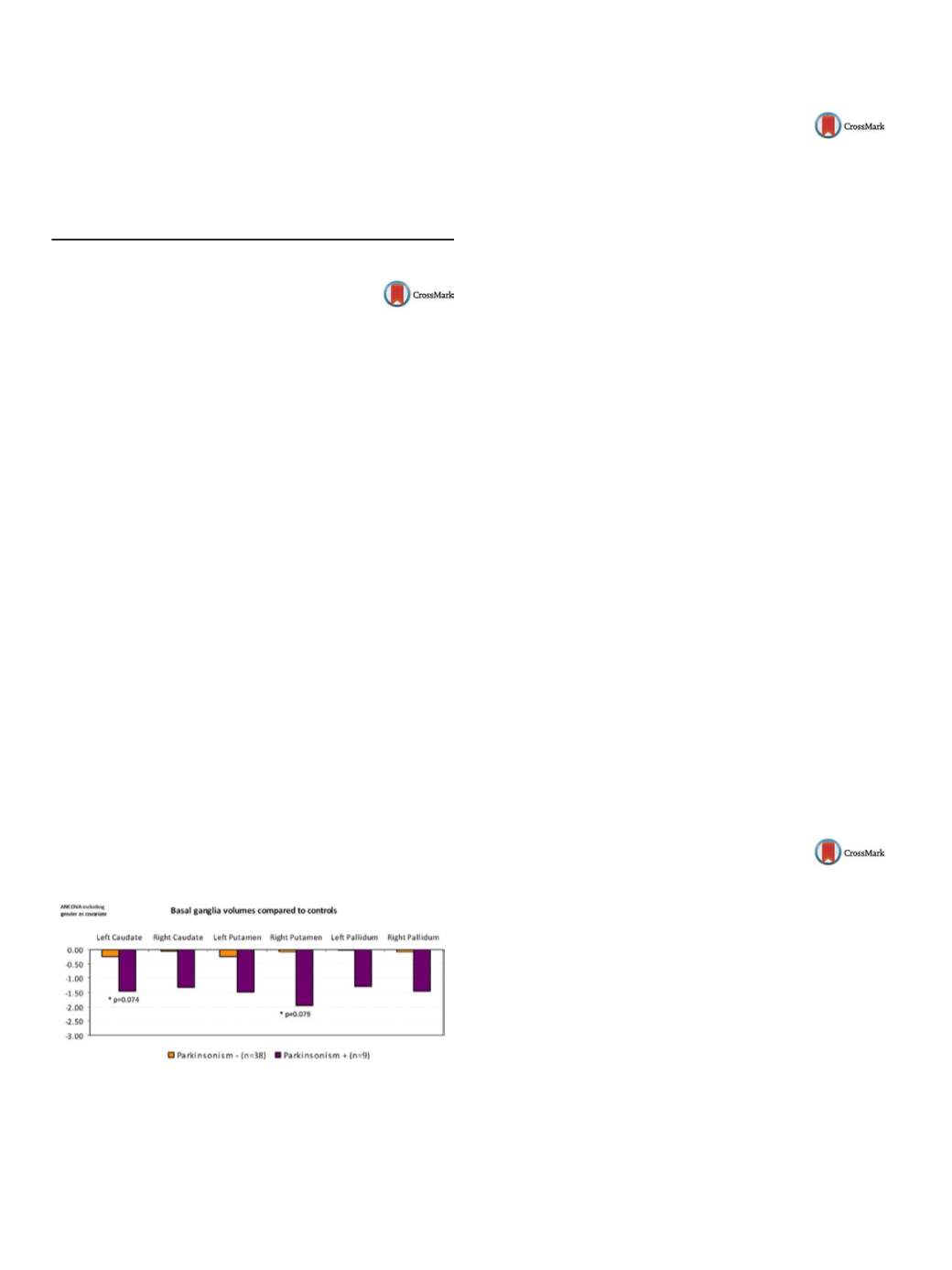
Results
Patients with Parkinsonism showed a trend towards sig-
nificance to exhibit reduced volumes in the left caudate and right
putamen
( Fig. 1 ).Conclusions
FEP patients who exhibit Parkinsonian signs tend to
show reduced left caudate and right putamen volumes in the early
phases of psychotic illness, after correcting by gender.
Fig. 1
Disclosure of interest
The authors have not supplied their decla-
ration of competing interest.
http://dx.doi.org/10.1016/j.eurpsy.2016.01.046FC43
The role of neurometabolites in
emotional processing
D. Denzel
1 ,∗
, L.R. Demenescu
2, L. Colic
2, F. von Düring
3,
H. Nießen
3, M. Walter
21
Magdeburg, Germany
2
Clinical Affective Neuroimaging Laboratory CANLAB, Leibniz
Institute for Neurobiology, Magdeburg, Germany
3
Clinical Affective Neuroimaging Laboratory CANLAB,
Otto-von-Guericke University Magdeburg, Magdeburg, Germany
∗
Corresponding author.
Objective
To investigate how brain metabolites, especially glu-
tamate and glutamate to glutamine ratio of pgACC modulate the
neural response within these areas and how this affects their func-
tion during emotion facial expression matching task.
Methods
Seventy healthy volunteers underwent magnetic reso-
nance spectroscopy (MRS) and task functional magnetic resonance
imaging (fMRI) in 7 Tesla scanner. PgACC MRS data were obtained
using STEAM sequence and analyzed using LCModel.
Angry, fearful, and happy facial expressions were presented in an
affect-matching block where one of the two facial expressions pre-
sented matched the target facial expression. The control condition
was form matching. Data were preprocessed and analyzed in SPM
8.
Results
Glutamate to Creatine ratiomeasured inpgACC positively
correlated with BOLD response in the right DLPFC during neg-
ative emotional perception (FWE = 0.05) Glutamate to glutamine
ratio indicating on-off mechanisms in pgACC positively corre-
lated with BOLD responses in FFA extending to cerebellum cluster
(FWE < 0.05).
Conclusion
This study indicate that pgACC, baseline metabolism
predicts neural response to emotional
processing.Weconclude that
individuals with higher glutamate ratios, an excitatory neurotrans-
mitter, in pgACC during rest might have a better copingmechanism
to potential danger indicated by perception of angry or afraid faces.
The higher glutamate to glutamine ratio in pgACC indicates a higher
turnover of excitatory metabolite glutamate. This mechanism is
associated with higher emotional response in fusiform area and
cerebellum suggesting higher visual attention towards negative
emotions.
Disclosure of interest
The authors have not supplied their decla-
ration of competing interest.
http://dx.doi.org/10.1016/j.eurpsy.2016.01.047FC44
Association analysis of imbalanced
interhemispheric functional
coordination and early therapeutic
efficacy in major depressive disorder:
Evidence from resting state fMRI
Z. Hou
1 ,∗
, X. Song
2, W. Jiang
1, Y. Yue
1, Y. Yin
1, Y. Zhang
1,
Y. Liu
3, Y. Yuan
11
Affiliated Zhongda Hospital of Southeast University, Medical School
of Southeast University, Department of Psychosomatics and
Psychiatry, Nanjing, China
2
College of Engineering, Peking University, Department of
Biomedical Engineering, Beijing, China
3
Key Laboratory of Cognition and Personality, Faculty of Psychology,
Southwest University, Chongqing, China
∗
Corresponding author.
Introduction
Emerging evidences indicate that the alteration of
interhemispheric functional coordination may be involved in the
pathogenesis of major depressive disorder (MDD). In present study,
we aimto explore the potential marker by using the voxel-mirrored
homotopic connectivity (VMHC) approach, which may be con-
tributing to predict the clinical prognosis in MDD.