


 237 / 812

24th European Congress of Psychiatry / European Psychiatry 33S (2016) S116–S348
S233
Methods
After obtaining ethical clearance from the Institute
Review Board, we recruited 30 subjects with active mania and 15
healthy controls using purposive sampling. Computerized version
of the Stroop Colour Word Test was used.
Results
The two groups were similar on socio-demographic vari-
ables. No difference in performance was seen in the two groups on
the total number of correct and incorrect responses and reaction
times for correct responses on incongruent condition of the Stroop
Test. However, subjects in the mania group were quicker inmaking
incorrect responses. During incongruent condition of Stroop test, in
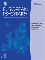
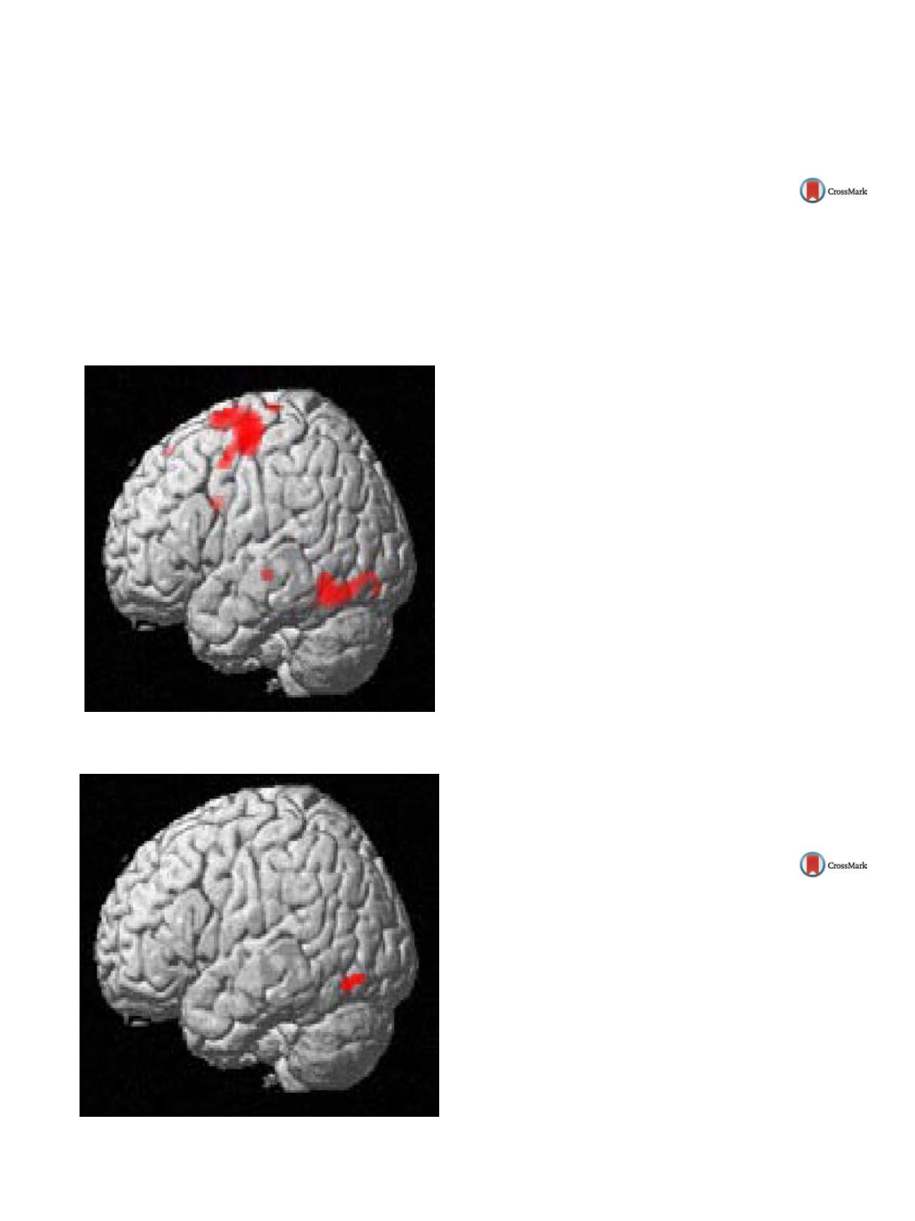
the mania group
( Fig. 2 ),only left fusiform gyrus was activated in
comparison to the control group
( Fig. 1 ),which had left cingulate
gyrus, right frontal lobe, and superior temporal gyrus activation.
Discussion
It is evident that mania group performed relatively
poorly on response inhibition task since they took lesser time
to make incorrect responses. This may be explained by the non-
activation of frontal areas, responsible for the executive functioning.
Fig. 1
Fig. 2
Disclosure of interest
The authors have not supplied their decla-
ration of competing interest.
http://dx.doi.org/10.1016/j.eurpsy.2016.01.461EW344
Decreased interhemispheric
functional coordination underlying
the cognitive impairment in
late-onset depression
Z. Hou
1 ,∗
, W. Jiang
1, Y. Yue
1, Y. Yin
1, Y. Zhang
1, Y. Sui
2,
Y. Yuan
11
Affiliated Zhongda Hospital of Southeast University, Medical School
of Southeast University, Psychiatry, Nanjing, China
2
Affiliated Nanjing Brain Hospital of Nanjing Medical University,
Psychiatry, Nanjing, China
∗
Corresponding author.
Background
The intuitive association between cognitive dys-
function in late onset depression (LOD) and the aberrant functional
activity in the brain’s default-mode network (DMN) has prompted
interest in exploring the role of the DMN in LOD. The altered pattern
of resting state voxel-mirrored homotopic connectivity (VMHC) in
cognitive processes is not yet well understood in LOD.
Methods
The study was designed to examine the implicit
coupling between the alteration of interhemispheric functional
coordination and cognitive impairment in LOD. Thirty-one LOD
patients and 37 matched healthy controls (HC) underwent neu-
ropsychological tests and functional magnetic resonance imaging
(fMRI) in this study.
Results
Compared to HC group, attenuated VMHC in superior
frontal gyrus, superior temporal gyrus, posterior cerebellar lobe,
postcentral and precentral gyrus was observed in LOD. Neuro-
behavioral relevancy approach revealed that the imbalanced
interhemispheric functional coordination in bilateral cerebellum
was positively correlated with the performance of trail making test
in LOD (
r
= 0.367,
P
= 0.040).
Conclusion
Altered linkage pattern of intrinsic homotopic con-
nectivity and cognitionwas firstly investigated in LOD, and it would
provide a novel clue to reveal the neural substrates underlying the
cognitive dysfunction in LOD.
Keywords
Late-onset depression; Voxel-mirrored homotopic
connectivity; Functional magnetic resonance imaging; Cognitive
function; Cerebellum
Disclosure of interest
The authors have not supplied their decla-
ration of competing interest.
http://dx.doi.org/10.1016/j.eurpsy.2016.01.462EW345
qEEG correlates of induced anxiety in
obsessive-compulsive patients –
comparison of autobiographic and
general anxiety
D. Kamaradova
∗
, J. Prasko , K. Latalova , A. Grambal , J. Taborsky ,
M. Hajda
University Hospital Olomouc, Psychiatry, Olomouc, Czech Republic
∗
Corresponding author.
Introduction
Obsessive-compulsive disorder (OCD) is character-
ized by the presence intrusive thoughts (obsessions) that cause
anxiety in patients. Patients then perform various types of rituals
(compulsions) to suppress symptoms of anxiety. OCD differ from
other anxiety disorders. In OCD patients, the anxiety is caused by
individual’s specific situation. Aimof our studywas to compare EEG
signal during resting state, authobiographic scenario and general
anxiety scenario.
Methods
Resting-state eyes-closed EEG data were recorded in
twenty OCD patients and fifteen healthy controls that were